Test tube rack to hold one small and one
large diameter test tube, for a hospital laboratory. The surface should be smooth and easy to clean,
and resistant to ethanol and acetone. The rack should be sufficiently heavy to resist
being knocked over, and sturdy enough to survive being dropped on the floor.
The purpose of this test tube rack is to
provide a hands free method of holding the
tube upright. There are two important reasons:
1.
To enable laboratory staff to fill the tube
with liquid expelled from a syringe. Two hands are needed when operating a
syringe.
2.
Not holding the test tube with
a hand, reduces the risk of accidental needle stick injury to the laboratory
staff when filling the test tube from a syringe with a needle.
Background: The types of specimens
collected in these test tubes is material sucked out of patients lumps, with a
needle and a syringe. A cytology scientist prepares the liquid specimens into
glass slides. I am a medical doctor trained as a Pathologist. I work in a hospital
laboratory. I examine the patient’s lump, suck out some fluid with a needle and
syringe, collect the specimen in the
test tube, and look down the microscope
to make the diagnosis. The diagnosis is the explanation as to what is causing
the lump, examples are cancer or infection.


The
laboratory scientist is expelling liquid from a pipette into the test tube.
This larger rack holds up to 8 test tubes, and is used on the laboratory bench.
We need a smaller test tube rack to take out when collecting samples from
patients. The smaller rack is what you are designing. In a hospital laboratory we wear gloves to
prevent contact with infectious material and chemicals. These purple gloves are
resistant to acetone.

This
is Dr Low, she is a Pathologist, she is looking down the microscope at a glass
slide, to make a diagnosis. She is in charge of Cytology specimens, which is
the area we need the test tube rack.
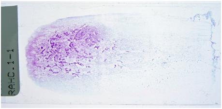

On the top is a glass slide with cells,
stained purple so we can see them. Below it is what the cells look like
down the microscope, the magnification is 400x what you can see with your eye.

This is me, Amanda Charlton, I work in this
office in the Histology Department of Middlemore Hospital in Auckland. I use a microscope
to look at samples, and I voice dictate my diagnostic reports onto the computer.
See the microscope has an extra arm to the left side, this is to allow a second
person to look down and see the same thing I am seeing. This is called a double
header microscope, and it is used for teaching young doctors to be
pathologists.
No comments:
Post a Comment